From grandmothers down to grandoffspring: grandchicks of old grandmas are born “old”

As animals age, they generally look less good and their telomeres, small structures that protect chromosomes from becoming frayed or tangled, become shorter. In our article we investigated if the effects of age at conception of mothers to the telomeres of their offspring would persist over a subsequent generation (grandoffspring generation), having previously found out that they persisted into the offspring generation.

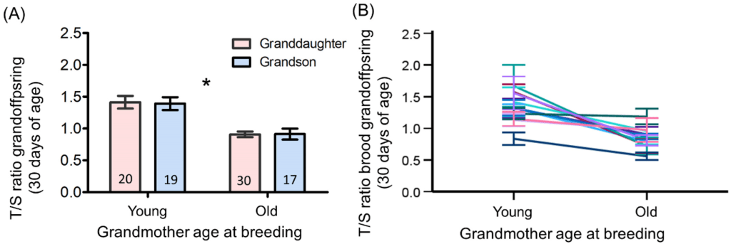
In this new study, we showed that the shortened telomeres found in the joungsters of older grandmothers are also present in their children, i.e. the grandchildren generation – even if the breeding mothers of the 2nd generation were young. This effect was considerable: telomeres were 43% shorter in the offspring of grandmothers who were old at rearing than in the offspring of the same grandmothers who were young at rearing. Shorter telomeres at the time of fledging are associated with a shorter lifespan in zebra finches. These results indicate that it is necessary to look beyond a single generation to explain inter-individual differences in ageing and different age-specific reproductive efforts. The mothers were young at the time of breeding, so effects due to the age of these mothers can be ruled out. It would be very interesting to know whether the effects of the grandmother’s age increase if the mother’s age is also high.

Our data reveal a hidden legacy that can be passed on across generations and has a negative impact on the lifespan and reproductive value of offspring. We stress, therefore, that evolutionary biologists and ecophysiologists need to look beyond a single generation and current environmental conditions to fully understand the causes of inter-individual differences in ageing rates and age-specific reproductive effort.

The article by Valeria Marasco, Winnie Boner, Kate Griffiths, Shirley Raveh and Pat Monaghan is accessible Open Access 🙂

We are very much grateful to the PR team at Vetmeduni for helping us to communicate our work to broader audiences and contribute to our #OutreachMission #OpenScience Vetmeduni PR article (in German and in English). 

From Transcriptomics to Seasonality: brain gene expression reveals how birds may time their nocturnal migrations

Drawn by Zuzanna Zielinska

How do birds know when it is time to start their nocturnal long-distance migratory flights? Or, how do mammals know when it is time to hibernate? How modulation in physiology is regulated has fascinated, as much as puzzled, researchers since centuries. It is relatively well understood that the timing of migration in birds is activated by changes in photoperiod as well as internal changes. However, the molecular underpinnings that allow these signals to change seasonally and daily is largely unknown. Filling this knowledge gap is critical to understand how seasonality has driven the evolution of extreme physiological life-history strategies, such as migration and hibernation.

In our new study, we exposed young adult quail (Coturnix coturnix) to controlled changes in day length to simulate autumn migration, and then blocked the photoperiod until the birds entered the non-migratory overwintering phase. We then carried out RNA sequencing of selected brain samples (hypothalamus) taken from birds at a standardized time during the night when the birds entered a “restlessness state” (which is a robust proxy of readiness to migrate in the lab) and their body mass reached the peak.

We found that such restlessness state was linked to an upregulation of a few, but functionally well-defined gene expression networks involved in fat transport, protein and carbohydrate metabolism. We then performed further experiments focusing on two candidate genes (apolipoprotein H [APOH], lysosomal-associated membrane protein-2 [LAMP2]) from day-time and night-time samples from the entire study population. Such analyses revealed differences in the expression of these genes depending on the time of day, with the highest expression levels found in samples from migratory birds taken at night. We also found that the expression of APOH in migratory birds was positively associated with nocturnal activity, while such association was absent within the non-migratory birds.

The results provide novel experimental evidence that hypothalamic changes in the expression of apolipoproteins, which regulate the circulating transport of lipids, are likely to be key regulatory activators of nocturnal migratory movements. We hope that our study will stimulate further functional investigations of the seasonal physiological remodelling underlying the development of the migratory phenotype. Understanding the neurophysiological substrates by which highly seasonal species such as migratory and hibernating vertebrates can adjust their energy metabolism seasonally and daily is key if we are to understand the impact of ongoing climatic challenges on the life history and fitness of organisms as we enter the disruptive phases of the Anthropocene.

The article “Brain gene expression reveals pathways underlying nocturnal migratory restlessness“ by Valeria Marasco, Leonida Fusani, Patricia Haubensak, Gianni Pola and Steve Smith is accessible Open Access 🙂

We are very much grateful to the PR team at Vetmeduni for helping us to communicate our work to broader audiences and contribute to our #OutreachMission #OpenScience Vetmeduni PR article (in German). A big thank goes to Zuzanna Zielinska who made the beautiful illustration of the flying male and female quail (above).

In preparation for migration, birds accumulate fat stores and modulate their innate immune functions, our latest research suggest

In preparation for migration, common quail accumulate fat stores and modulate their innate immune functions. They increase their immunity midway through the fattening process. Individuals that accumulate less fat maintain a higher immune response, suggesting a competitive relationship between these processes, explored in the paper by Marcin Tobolka and colleagues. Image credit: Zuzanna Zielinska. Creative Commons Attribution 4.0 International license.

During their migrations, migratory birds are exposed to numerous pathogens. A functioning immune system is therefore crucial when flying into new environments. However, immunity is energetically costly and competes with other important physiological processes. In our latest work, we examined the extent to which fattening before bird migration influences innate immunity using the common quail as our study species.

While many studies showed that the immunity of birds is often weakened during their strenuous migratory flights, it remains little understood if and how immunity changes during the rapid accumulation of energy stores in preparation for migration. To fill this knowledge gap, we induced pre-migratory fattening through controlled changes in daylight and regularly examined changes in three markers of innate immunity – leukocyte coping capacity (LCC), hemagglutination and hemolysis titers. In addition, body composition, namely lean mass and fat mass, was measured.

We found that all three markers showed similar changes during pre-migratory fattening. The LCC responses, hemagglutination and hemolysis titers were on average higher in the middle fattening phase than in the high fattening phase, here the values ​​were similarly high as before the start of pre-migratory fattening. In the middle phase of fattening, we found that the birds that had a higher fat content had lower peak LCC values ​​and hemolysis titers. Conversely, at this time of fattening, the birds with a higher proportion of lean mass had the highest peak LCC values. We proposed that these results could be due to competing or opposing processes between metabolic remodeling and the function of the innate immune system.

These three immune indicators are integral parts of the innate immune system, which acts quickly and effectively against a wide range of pathogens, even without prior exposure. This aspect is crucial in the context of animal migration, as the ability to respond quickly to various threats is crucial for fitness. For the future, we hope that our work will encourage further studies to obtain detailed information about the remodelling of the immune system during the most energetically demanding phases in the life cycle of migratory birds.

Our article “Controlled expression of avian pre-migratory fattening influences indices of innate immunity” by Marcin Tobolka, Zuzanna Zielińska, Leonida Fusani, Nikolaus Huber, Ivan Maggini, Gianni Pola, Valeria Marasco Biol Open 15 January 2024; 13 (1): bio060018. doi: https://doi.org/10.1242/bio.060018 is freely accessible and open access.

We are massively grateful to the PR team at Vetmeduni for helping us to communicate our work to broader audiences: https://www.vetmeduni.ac.at/klivv/ueber-uns/aktuelles

How do birds regulate seasonal expression of the migratory state? The gut hormone ghrelin could play a key role, suggests an experimental study in common quails

The seasonal expression and the migratory phenotype is signalled by rapid and marked changes in food intake, fuelling, and amount of nocturnality. We used common quails (Coturnix coturnix) to assess if the hormone corticosterone and the gut-derived hormone ghrelin play a role in these astonishing physiological transitions. We exposed quails to controlled changes in day length to simulate autumn migration, followed by a wintering period. We compared corticosterone and ghrelin concentrations and assessed whether these two metabolic hormones varied between distinct migratory states. We found that the expression of the migratory phenotype was associated with a sharp elevation in circulating concentrations of ghrelin. Interestingly, we also found that concentrations of ghrelin correlated with changes in body mass (fat stores) of birds as they transitioned into their autumnal migratory state and as they entered the wintering state. Contrary to our predictions we did not detect functional links between plasma corticosterone and the physiological state of the birds and we also did not observe a link between circulating levels of ghrelin and corticosterone.

The article “Ghrelin, not corticosterone, is associated with transitioning of phenotypic states in a migratory Galliform” by Valeria Marasco, Hiroyuki Kaiya, Gianni Pola & Leonida Fusani can be accessed open access here. Vetmeduni Press Release.

What to measure to predict brain size in birds?

At least in part, the brain’s capacity to process cognitive processes depends on the mass of neural tissue involved – the more tissue, the more information can be processed. Studies often find a positive relationship between brain size and cognitive performance. However, majority of these studies are based on comparisons between different species. Scientists are now trying to understand how more subtle differences between individuals of the same species are related to their cognitive skills, which is often a big challenge when studying animals in nature.  

A first study in the barn swallow proposed to use external head measurements, which require handling but not the sacrifice of the study subject, as an accurate approximation for brain mass. In this collaborative research with Joanna Bialas and Marcin Tobółka (Poznań University, Poland), we employed this method for the first time in a small Galliform, the Common Quail. We measured both the external head dimensions of the birds as well as the weight of their brains, and tested how well these two measurements were related to each other. Despite we did find that these measurements were correlated, the correlation values we found were not strong enough to allow using external head measurements to predict an individual’s brain mass with high confidence. Moreover, the best predictor of brain mass was not head volume, as previously demonstrated in barn swallows, but the height of the head alone. We therefore recommend validating the original method of external head measurements in each species before making assumptions on how these measurements might be related to brain size and cognitive performance.  

Photo credit: Stefan Graf

You can access a full version of the article here

The work was funded by the FWF Der Wissenschaftsfonds Lise Meitner Fellowship (M2520-B29)

Stress influences telomere dynamics as predicted by changes in mortality rates

It is a pleasure to introduce our last paper on stress and ageing in #theFASEBJournal

This latest paper is associated with our previous findings where we found that the repeated exposure to challenging environmental circumstances during adult life increased chances of survival during middle adulthood, but not during young- or old-adulthood. Here we report that these effects are underpinned by changes in telomere dynamics, with stress-exposed birds showing reduced telomere loss in middle adulthood. Our data provides novel experimental evidence that telomere dynamics play a key role linking stress resilience and aging.

Avian fitness: birds go easy on their pectoral muscles

PressRelease by the Vetmed about our freshly published article in Journal of Experimental Biology:

“Controlled expression of the migratory phenotype affects oxidative status in birds”

by myself, Manrico Sebastiano, David Costantini, Gianni Pola, Leonida Fusani

Really proud to see this out. A big challenge since the very beginning. It “officially” all started in 2017 in an empty room in the Konrak Lorenz Institute of Ethology (Vetmeduni Vienna) with 0 Common quails 🙂

Link to access the full article: https://jeb.biologists.org/content/224/6/jeb233486

Transcriptome of the migratory Common quail

Check out our freshly published open access paper on the brain de novo transcriptome of the migratory Common quail in Data in Brief:

In this article, we developed a high quality pipeline for compiling transcriptomes of complex non-genomic species. Our data not only can provide a robust reference for gene expression studies in migratory birds but will also be useful for studies in related Galliforms, including the Japanese quail, a recognised model in behavioural neuroendocrinology and physiology research.

quaglia_in_canto_GianniPola

@Photo credit: Gianni Pola